Safety Precautions
Critical Safety Requirements:
- Laser Safety: CytoFLEX contains Class 3B lasers. Never defeat safety interlocks or look directly into laser beam paths.
- Biological Hazards: All samples must be treated as potentially infectious. Use BSL-2 practices minimum.
- Chemical Safety: Cleaning solutions and reagents may be hazardous. Review all Safety Data Sheets.
- Electrical Safety: Ensure proper grounding and electrical safety compliance.
- Waste Disposal: Follow institutional guidelines for disposal of biohazardous and chemical waste.
Introduction to Flow Cytometry
Flow cytometry is a powerful analytical technique that allows for the rapid analysis of physical and chemical characteristics of particles or cells as they flow in a fluid stream through a beam of light. Beckman Coulter's CytoFLEX and Navios systems represent state-of-the-art flow cytometry technology, providing researchers and clinical laboratories with precise, reliable cell analysis capabilities.
This comprehensive guide will introduce you to the fundamental concepts of flow cytometry, explain how Beckman Coulter systems work, and provide practical guidance for successful operation and basic troubleshooting.
Fundamental Principles of Flow Cytometry
Flow cytometry operates on several key principles that work together to analyze individual cells:
Hydrodynamic Focusing
The sample is injected into a flowing stream of sheath fluid, which narrows the sample stream so that cells pass through the laser interrogation point in single file. This hydrodynamic focusing ensures that each cell is individually analyzed and prevents cells from clustering together.
Light Scattering
When a cell passes through the laser beam, it scatters light in different directions:
- Forward Scatter (FSC): Correlates with cell size - larger cells scatter more light forward
- Side Scatter (SSC): Correlates with internal complexity - cells with more internal structures scatter more light to the side
Fluorescence Detection
Fluorescent labels (fluorochromes) attached to antibodies or other probes emit light when excited by the laser. Different fluorochromes emit at different wavelengths, allowing for simultaneous detection of multiple cellular characteristics.
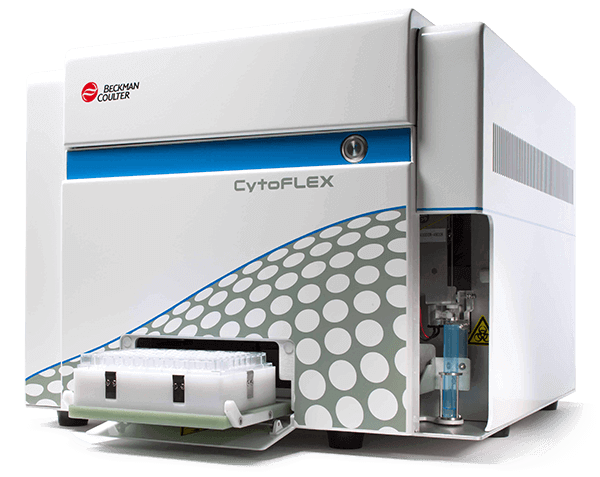
Beckman Coulter CytoFLEX System Overview
The CytoFLEX represents Beckman Coulter's newest generation of flow cytometers, designed for flexibility and ease of use:
Key Features
- Modular Design: Configurable with 1-4 lasers (405, 488, 561, 638 nm)
- Up to 21 Parameters: Simultaneous measurement of multiple characteristics
- Small Sample Volumes: As little as 12 μL per test
- Easy Maintenance: Daily QC in under 3 minutes
- CytExpert Software: Intuitive analysis and data management
System Components
- Fluidics System: Manages sample delivery and waste handling
- Optical System: Lasers, filters, and detectors for light detection
- Electronics: Signal processing and data conversion
- Software: User interface and data analysis tools
Sample Preparation Fundamentals
Proper sample preparation is critical for reliable flow cytometry results:
Cell Preparation Guidelines
- Cell Viability: Use fresh samples when possible. Cells should be >90% viable for optimal results.
- Cell Concentration: Adjust to 1-5 × 10⁶ cells/mL for most applications
- Single Cell Suspension: Filter through 40-70 μm mesh to remove clumps
- Buffer Selection: Use appropriate buffers (PBS, HBSS) with proper pH and osmolality
Antibody Staining Protocol
- Wash cells in staining buffer (PBS + 2% FBS + 0.09% sodium azide)
- Resuspend at appropriate concentration (usually 1 × 10⁶ cells/100 μL)
- Add antibodies at manufacturer-recommended concentrations
- Incubate in dark at 4°C for 15-30 minutes
- Wash twice to remove unbound antibodies
- Resuspend in analysis buffer
Basic System Operation
Operating a CytoFLEX system follows a standardized workflow:
Daily Startup Procedure
- System Preparation (5 minutes):
- Turn on system and allow lasers to warm up
- Check fluid levels (sheath, cleaning solution, waste)
- Verify environmental conditions are stable
- Quality Control (3 minutes):
- Run CytoFLEX Daily QC using provided beads
- Verify all parameters are within acceptable ranges
- Save QC results for documentation
- Optical Alignment Check:
- Use alignment beads to verify laser alignment
- Adjust if necessary following manufacturer procedures
- Document any adjustments made
Sample Analysis Workflow
- Sample Loading: Place prepared sample in sample tube holder
- Acquisition Setup: Configure parameters, gates, and collection criteria
- Threshold Settings: Set appropriate FSC and SSC thresholds to exclude debris
- Compensation: Apply fluorescence compensation for spectral overlap
- Data Collection: Acquire sufficient events (typically 10,000-100,000)
- Data Analysis: Use gating strategies to identify cell populations of interest
Understanding Flow Cytometry Data
Flow cytometry generates complex data that requires proper interpretation:
Data Display Methods
- Dot Plots: Two-parameter displays showing correlation between measurements
- Histograms: Single-parameter distributions showing population frequencies
- Contour Plots: Population density representations
- Overlay Plots: Comparison of multiple samples or conditions
Gating Strategies
Gating allows you to select specific cell populations for analysis:
- Primary Gate: Usually FSC vs SSC to select viable cells
- Secondary Gates: Additional parameters to refine population selection
- Boolean Gates: Combine multiple parameters using AND/OR logic
- Time Gates: Exclude events from unstable acquisition periods
Common Applications
Flow cytometry has numerous applications across research and clinical fields:
Clinical Applications
- Immunophenotyping: Characterizing cell surface markers
- Cell Cycle Analysis: Determining DNA content and proliferation
- Apoptosis Detection: Measuring programmed cell death
- Intracellular Cytokines: Detecting internal protein expression
Research Applications
- Stem Cell Analysis: Identifying and sorting stem cell populations
- Cancer Research: Studying tumor cell characteristics
- Drug Discovery: Screening compound effects on cells
- Environmental Monitoring: Analyzing microbial populations
Basic Troubleshooting
Understanding common issues helps ensure reliable results:
Poor Data Quality
Symptoms: High background, poor resolution, excessive debris
Solutions:
- Check sample preparation quality
- Verify proper antibody concentrations
- Ensure adequate washing steps
- Filter samples to remove aggregates
- Check for proper storage conditions
System Performance Issues
Symptoms: Unstable flow rates, poor QC results, laser fluctuations
Solutions:
- Run system cleaning cycles
- Check fluidics for blockages or bubbles
- Verify sheath fluid quality and levels
- Inspect sample lines for contamination
- Contact service if laser performance degrades
Maintenance and Care
Regular maintenance ensures optimal system performance:
Daily Maintenance
- Run daily QC before each use
- Clean sample lines after each session
- Check and refill fluid reservoirs
- Empty waste containers when 80% full
- Wipe down external surfaces
Weekly Maintenance
- Deep clean fluidics system
- Replace filters if indicated
- Check optical alignment
- Verify environmental conditions
- Update maintenance logs
Safety Considerations
Flow cytometry involves several safety considerations:
Laser Safety
- Never look directly into laser beams
- Use proper laser safety glasses when required
- Ensure safety interlocks are functional
- Post appropriate warning signs
Biological Safety
- Treat all samples as potentially infectious
- Use appropriate personal protective equipment
- Follow institutional biosafety guidelines
- Properly dispose of biological waste
| Parameter | Light Source | Detection | Information Provided |
|---|---|---|---|
| Forward Scatter (FSC) | 488 nm laser | Forward detector | Cell size |
| Side Scatter (SSC) | 488 nm laser | 90° detector | Cell granularity/complexity |
| FITC/Alexa Fluor 488 | 488 nm laser | 525/40 filter | Green fluorescence |
| PE | 488 nm laser | 585/42 filter | Orange fluorescence |
| APC | 638 nm laser | 660/10 filter | Red fluorescence |
| DAPI/Hoechst | 405 nm laser | 450/45 filter | DNA staining |